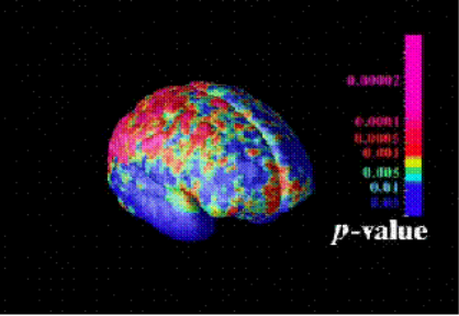
The Schizophrenic Brain... (see images below)
Cast thy burden upon the Lord, and he shall sustain thee. Psalm 55:22
I added this more comprehensive link because I found that my links on this topic kept "disappearing"... so, you may just want to go to this new link to read the whole article... but, before you do that, I want you to note the comment on the "NON-GENETIC" trigger in the article below... it is highlighted in red for you. This is in a shorter article that also discusses the tremendous gray matter loss in schizophrenia... another article from the NIMH (National Institute of Mental Health). Again, note the comment regarding "NON-GENETIC" trigger before leaving this page... there are also pictures below you may want to look at... so, I encourage you to look at all this information... as hard as that is to do!
MORE COMPREHENSIVE ARTICLE - MORE PICTURES TOO - CLICK HERE!
STARTS HERE - WORD FOR WORD QUOTE FROM: http://www.nimh.nih.gov/events/prteens.cfm
"National Institute Of Mental Health NEWS UPDATE
Teens With Schizophrenia Lose Gray Matter in Back-to-Front Wave
Contact: Jules Asher
301-443-4536
E-mail: nimhpress@nih.gov
Brains of teens with early onset schizophrenia are ravaged by a back-to-front wave of gray matter loss that parallels the progression from hallucinations and delusions to thinking and emotional deficits, National Institute of Mental Health (NIMH) - supported scientists have discovered. This loss of critical working brain tissue begins in rear perception processing areas, and over 5 years engulfs frontal areas responsible for functions like planning and reasoning. Although some loss of neurons and their branch-like extensions is normal during the teen years, as the brain prunes unused connections, the researchers had earlier shown that teens with childhood onset schizophrenia lose 4 times the normal amount in their frontal lobes. The new study is the first to visualize such a pattern of progressive tissue loss in schizophrenia. Paul Thompson, M.D., University of California, Los Angeles (UCLA), Judith Rapoport, M.D., NIMH, and colleagues, report on their findings in the September 25, 2001, Proceedings of the National Academy of Sciences.
Using magnetic resonance imaging (MRI), the researchers periodically scanned 12 teens with schizophrenia and 12 age-matched healthy teens over 5 years, beginning at age 14. The wave of gray matter loss began in an area above the ear and then spread forward. Since losses in the rear areas are thought to be caused by environmental factors, the findings are consistent with the notion that activation of some non-genetic trigger contributes to the onset and initial progression of the illness, suggest the researchers. The wave of loss correlated with worsening psychotic symptoms and mirrored the progression of neurological and cognitive deficits associated with the disorder. The final profile was consistent with the loss pattern in adult schizophrenia. Another group of 10 teens who were taking anti-psychotic medications for a different disorder did not show the same pattern of changes, reducing the likelihood that the gray matter losses were drug-induced.
The study is part of ongoing research on childhood onset schizophrenia by the NIMH Child Psychiatry Branch, which Rapoport heads. It employed a new 3-D MRI image analysis technique developed by NIMH grantee Arthur Toga, M.D, UCLA Laboratory of
Neuroimaging, and colleagues. Support was also provided by grants from the NIH National Center for Research Resources, National Institute on Neurological Disorders and Stroke, and National Institute on Drug Abuse.
Also participating in the study were Jay Giedd, M.D., Peter Gochman, Jonathan Blumenthal, Robert Nicolson, M.D., NIMH, and Toga and Christine Vidal, UCLA.
The National Institute of Mental Health (NIMH) is part of the National Institutes of Health (NIH), the Federal Government's primary agency for biomedical and behavioral research. NIH is a component of the U.S. Department of Health and Human Services.
Early and Late Gray Matter Deficits in Schizophrenia
Areas of gray matter loss - shown in red and yellow -- spread from back-to-front (right to left) over 5 years in composite MRI scan data from 12 teens with childhood onset schizophrenia, beginning at age 14 (left). Red and yellow denotes areas of greater loss.
Movie: Schizophrenia Ravages Teen Brain
Rate of gray matter loss
Composite MRI scan data showing areas of gray matter loss over 5 years, comparing 12 normal teens (left) and 12 teens with childhood onset schizophrenia. Red and yellow denotes areas of greater loss. Front of brain is at left.
Source: Paul Thompson, M.D., UCLA, Laboratory of Neuroimaging
Posted: November 08, 2001
| NIMH Home | Welcome | News and Events | Clinical Trials | Funding Opportunities |
| For the Public | For Practitioners | For Researchers | Intramural Research |
| Top |
For information about NIMH and its programs, please email, write or phone us.
NIMH Public Inquiries
6001 Executive Boulevard, Rm. 8184, MSC 9663
Bethesda, MD 20892-9663 U.S.A.
Voice (301) 443-4513; Fax (301) 443-4279
TTY (301) 443-8431
Se Habla Español
[Disclaimer, Copyright, and Privacy Notice]
National Institutes of Health (NIH)
9000 Rockville Pike
Bethesda, Maryland 20892
Department of Health
and Human Services
END OF QUOTE/NIMH ARTICLE...
Schizophrenic brain images below are taken from: http://www.nimh.nih.gov/events/teenbrainvideo.cfm (click on "animation", then click on the picture of the brain to view video stream of damage to brain)...
The following are "snapshots" taken from that video... just to give you an idea... given the last time I looked, the video itself has "disappeared" from the NIMH website... this shows gray matter loss in persons with schizophrenia with puberty onset (MRI's were taken starting at age 14... you can already see damage there - a normal brain was shown as "blue")... second image below is what we see after 5 years... tremendous gray matter loss...
This study was published in September 25, 2001 Proceedings of the National Academy of Sciences. Things tend to "disappear" from the Internet. As such, I try to find "alternate links" with the same info. If the above link does not work, try one of these: http://www.loni.ucla.edu/~thompson/MEDIA/PNAS/NIMH.html, http://pn.psychiatryonline.org/cgi/content/full/36/22/18,Note: The animation provided on the above link shows loss of gray matter in schizophrenic teenagers (indicated by pink area). Note that the wave of degeneration occurs from back to front and that it obviously had already started prior to this imaging study - clearly already close to 1/3 of the brain is already impacted by age 14. This study was conducted over 5 years - so it ended around age 19 or 20! So, this loss of gray matter appears to take place over a period of 8 - 10 years.
The following provides a "slice" view of the schizophrenia brain: http://www.hcnr.med.harvard.edu/b_imaging/neurodeg/shizophrenia.html
Also note that the cerebellum is known to be involved in the coordination/regulation of motor functions, higher thought processes, emotions, and language. If the cerebellum (the "brain within the brain") regulates all these things, it stands to reason that it would be involved in the "reorganization" and "pruning" of the brain that occurs just before puberty.
The cerebellum is located in the back of the head, just above the brain stem. The spurt in "gray matter" thickening we normally see just prior to the onset of puberty starts from the back of the head and moves to the front.
It is a known fact that the cerebellum is smaller in the autistic child than in the normal child. IN my opinion, mercury may be lodging itself in the fragile, immature cerebellum area. It is a known fact that the cerebellum continues to mature until the early 20s. It is my THEORY that when the pre-puberty growth spurt in gray matter and the associated brain "reorganization/pruning" occur, that the mercury may "flow along" for the ride, thereby creating not "gray matter growth/thickening" (as you would normally see), but rather, dissolving the gray matter as it "flows along". Of course, that is just "my THEORY" as to what is going on, but in my opinion, it is a very probable one... and it certainly would explain why we see the damage we do in schizophrenia and why schizophrenia is diagnosed from puberty to age 40.
The schizophrenic brain loses 4X more gray matter at this stage of life than a normal brain. Note that persons who are schizophrenic today did not receive the compressed vaccination schedules that young autistic children have received. If this happens to persons who had less aggressive vaccination schedules, and mercury does play a part (and in my OPINION - it does), what will happen to autistic children as they reach this stage of life!!! :o(
Note: If I am correct in this theory, you would not see such devastation of gray matter in Alzheimer's since that "second wave growth spurt" occurs just before puberty and continues, apparently, for several years through adolescence. Keep in mind also that the cerebellum takes 20+ years to reach maturity - making it in my humble opinion a very, very fragile area indeed - and this is indeed one of the areas most impacted in autism! If mercury in flu shots, etc. are responsible for Alzheimer's, then that "second wave" would not occur in Alzheimer's. Hence this would explain why Autism and Alzheimer's appear to be MIRROR images in terms of "symptoms" as shown in the comparison of mental disorders. However, as the autistic child grows and hits pre-puberty and going forward, I very much suspect that his brain will begin to resemble that of the person with schizophrenia.
Note: 1 in 100 has Schizophrenia in the US! Schizophrenia is diagnosed anywhere from early teens to about age 40. Interestingly or - coincidently - mercury has a half life of 20 years... that would mean about 40 years for the body to "get rid of it". A schizophrenia diagnosis is rare after age 40. Parkinson's usually is diagnosed in the age 40 - 60 range. Alzheimer's hits primarily after age 65. In these disorders, are we seeing the "full life spectrum" of mercury damage? In my opinion, the answer to that is a resounding "YES"! Since this is a video animation, I had to do a screenprint and scan that - so, not the best quality image, but certainly enough to get the point across. This devastation, according to the NIMH, appeared over a 5 year period.
Note that even at the beginning of this experiment, already 1/3 of the brain had seen devastation... so, at what age does this start... 12? 10? 7? Children as young as 7 have been diagnosed with "childhood schizophrenia". Under that, they are usually called "autistic". So, if your child has made progress and then starts to regress, my guess would be that this is why! Again, this "teen" was not exposed to the compressed and more aggressive vaccination schedules young children have been exposed to either and as such, it truly makes me wonder - what will happen to our young children (now called "autistic") once they reach this age?
The NIMH website has removed the online video showing this change in "time lapse photography". It used to be found at: http://www.nimh.nih.gov/events/teenbrainvideo.cfm under an "animation" link.... in this screenprint below, you can see the actual link this came from: http://www.nimh.nih.gov/events/waveschizteen.avi
BLUE = normal , PINK = gray matter loss


Images Taken From: http://www.nimh.nih.gov/events/waveschizteens.avi
TO READ THE MUCH MORE COMPREHENSIVE ARTICLE - MORE PICTURES TOO - CLICK HERE!
Return To Autism-Schizophrenia-Alzheimer's Comparison Page
Cast thy burden upon the Lord, and he shall sustain thee. Psalm 55:22